68 (121)
6: Bacterial dermatoses
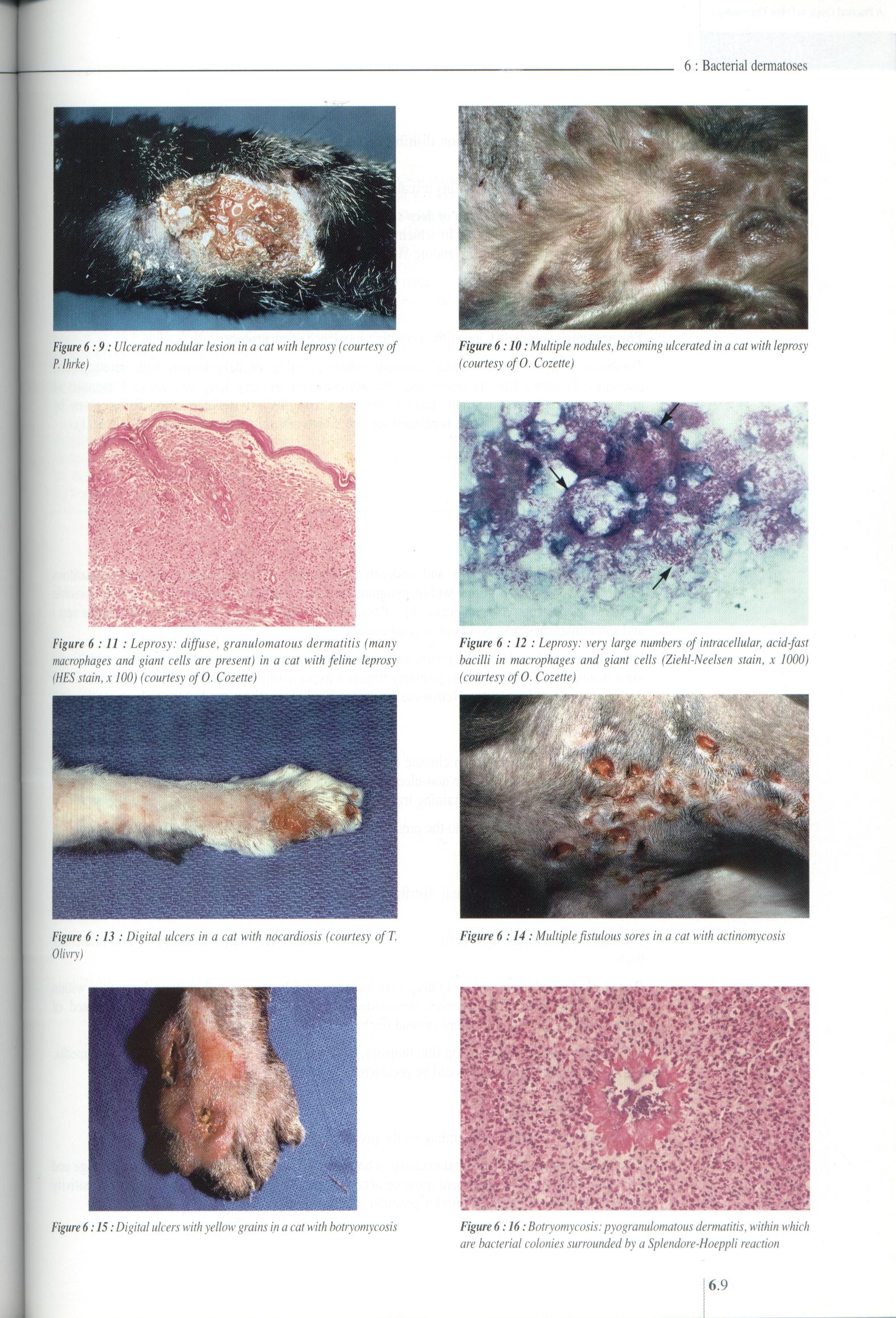
Figurę6:9: Ulceratednodular lesion ina cat with leprosy (courtesy of P.lhrke)
Figurę 6:10: Multiple nodules, becoming ulcerated in a cat with leprosy (courtesy of O. Cozette)
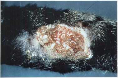
Figurę 6 :11 : Leprosy: diffuse, granulomatous dermatitis (many macrophages and giant cells are present) in a cat with feline leprosy (HES stain, x 100) (courtesy of O. Cozette)
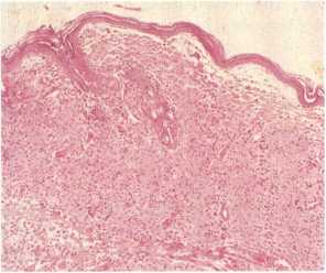
Figurę 6 :12 : Leprosy: very large numbers of intracellular, acid-fast bacilli in macrophages and giant cells (Ziehl-Neelsen stain, x 1000) (courtesy of O. Cozette)
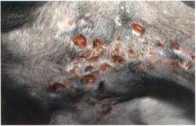
Figurę 6 :14: Multiple fistulous sores in a cat with actinomycosis
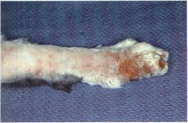
Figurę 6 :13 : Digital ulcers in a cat with nocardiosis (courtesy ofT. Olivry)
hm Wwtów
Z*
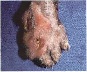
Figurę 6:15: Digital ulcers with yellow grains in a cat with botryomycosis
Figurę 6:16: Botryomycosis: pyogranulomatous dermatitis, within which are bacterial colonies surrounded by a Splendore-Hoeppli reaction
Wyszukiwarka
Podobne podstrony:
29 (352) 2: Diagnostic approach Figurę 2:17: Sclerosis in a cat with morphea (courtesy of E. Bensign
73 (103) 7 : Viral dermatoses Figurę 7:1: Ulcerative lesions on the upper and lower lip ofa cat with
64 (132) 6: Bacterial dermatoses Figurę 6:2: Secondarypyoderma in a cat with atopic dermatitis Figur
53 (169) 5 : Deep mycoses Figurę 5 :2 : Nasal nodule in a cat with phaeohyphomycosis Figurę 5:1: Ulc
55 (174) 5: Deep mycoses Figurę 5:9: Perinasal ulcer in a domestic short hair cal with cryptococcosi
74 (105) 7 A Practical Guide to Feline Dermatology ulcerated. Lesion distribution is multicentric bu
75 (101) 7: Viral dermatoses i■ft Figurę 7:9:Skinnecrosisofthepretemporalregion ina cat withfeline i
253 (15) 25 : Zoonotic dermatoses Figurę 25:1: Circular, well-defined, erythematous, inflammatory le
255 (16) 25 : Zoonotic dermatoses Figurę 25 : 9 : Nodular, inflammatory lesion on the back of the ha
61 (139) R. S. MuellerBacterial dermatoses Bacterial dermatoses, also called pyodermas, are rare in
62 (140) 6: Bacterial dermatoses Histopathological examination of skin biopsies may be consistent wi
66 (126) 6: Bacterial dermatoses antibiotics have been used in the treatment of atypical mycobacteri
77 (104) 7: Viral dermatoses Figurę 7:17: Epidermal horns on a metacarpal footpad ofa cat infected w
93 (100) 9 : Flea allergy dermatitis Figurę 9:1: Self-induced dorsolumbar alopecia in a Persian cal
95 (100) 9 : Flea allergy dermatitis Figurę 9:9: Extensive eosinophilic plaques in a cat with FAD Fi
43 (223) 4: Dermatophytosis Figurę 4:1: Erythematous blepharitis with comedones in a Persian cat wit
45 (221) Figurę4:9:Generalisedexfoliative dermatitis in a Persian cat with dermatophytosis caused by
47 (209) 4: Dermatophytosis Figurę 4 :17: Infected hairs in chloral lactophenol. Compare ectothrix i
b3cdb814f91df865 68/12) t nut dłwigm (71 68/121 *yi«£j nhajtą, Ł Odbapiaayi i wyj* **oml (ry«. (11/1
więcej podobnych podstron