SCAN0042

10 Clinical Anatomy of the Visual System
10 Clinical Anatomy of the Visual System
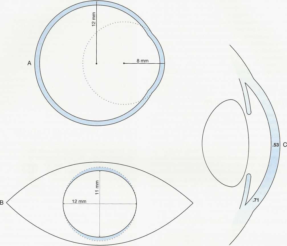
FIGURĘ 2-1
Corneal dimensions. A, Radius of curvature of cornea and selera. B, View from in front of the eye. The selera encroaches on the corneal periphery inferiorly and superiorly. Dotted lines show the extent of the cornea in the vertical dimension posteriorly. C, Sagittal section of cornea showing central and peripheral thickness (0.53 to 0.71 mm).
Wyszukiwarka
Podobne podstrony:
SCAN0131 96 Clinical Anatomy of the Visual System 96 Clinical Anatomy of the Visual System FIGURĘ 5-
SCAN0131 96 Clinical Anatomy of the Visual System 96 Clinical Anatomy of the Visual System FIGURĘ 5-
75024 SCAN0131 96 Clinical Anatomy of the Visual System 96 Clinical Anatomy of the Visual System FIG
SCAN0043 36 Clinical Anatomy of the Visual System FIGURĘ 3-2 Periphery of anterior segment of the gl
SCAN0044 38 Clinical Anatomy of the Visual System 38 Clinical Anatomy of the Visual System FIGURĘ 3-
86388 SCAN0154 264 Clinical Anatomy of the Visual System FIGURĘ 14-9 The near pupillary response. Do
33407 SCAN0128 90 Clinical Anatomy of the Visua! System 90 Clinical Anatomy of the Visua! System FIG
SCAN0033 crop Eye Axes Since the eye is not rotationally symmetric (i.e. the centers of curvature of
SCAN0033 crop Eye Axes Since the eye is not rotationally symmetric (i.e. the centers of curvature of
więcej podobnych podstron