SCAN0125

44 Clinical Anatomy of the Visual System
mented melanocytes, fibroblasts, and collagen bands1 (Figurę 3-12). The arrangement of these bands allows the ciliary body to slide against the selera without detaching from or stretching the tissue. The arrangement of the supraciliaris allows for the accumulation of fluid within its spaces, which may cause a displacement of the ciliary body from the selera. Damage to the layer caused by trauma may result in a ciliary body detachment.
CILIARY MUSCLE
The ciliary musde is composed of smooth muscle fibers oriented in longitudinal, radial, and circular directions. Interweaving occurs between fiber bundles and from layer to layer, such that varioirs amounts of connective tissue are found among the muscle bundles.1 The longitudinal muscle fibers (of Briicke) lie adjacent to the supraciliaris and parallel to the selera. Each muscle bundle resembles a long narrow V the base of which is at the ścierał spur, whereas the apex is in the choroid. The tendon of origin attaches the muscle fibers to the ścierał spur and to adjacent trabecular meshwork sheets. The insertion of the ciliary muscle is in the anterior one third of the choroid in the form of stellate-shaped terminations or "muscle stars."1,2 Below the longitudinal muscle fibers, the radial fibers form wider, shorter interdigitating Vs that originate at the ścierał spur and insert into the connective tissue near the base of the ciliary processes.1 This layer is a transition from the longitudinally oriented fibers to the circular fibers.
The innermost region of ciliary muscle, (Miiller's) annular muscle, is formed of circular muscle bundles with a sphincter-type action. These fibers are located near the major circle of the iris. Figurę 3-13 shows the relationship between these regions of the ciliary muscle and surrounding structures.
FSGURE 3-11 cont'd
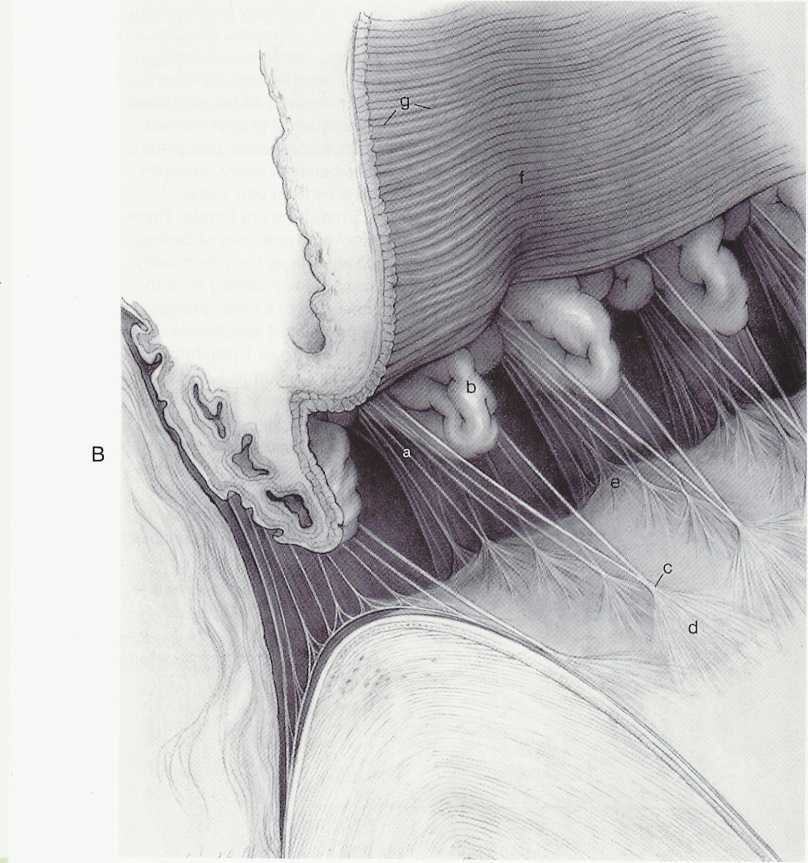
B, Anterior view of the ciliary processes showing zonules attaching to lens. Zonules form columns (a) on either side of ciliary processes (b), which meet on a single site (c) as they attach to lens. These two columns form a triangle, base of which is on ciliary body and apex of which is on lens. Zonules form tentlike structure (d) as they become attached to lens capsule. Equatorial surface of lens is crenated (e) by attachment of zonule. Iris is pulled upward, revealing its posterior surface containing radial folds (f) and circular furrows (g). (From Hogan MJ, Alvarado |A, Weddell JE: Histology of the human eye, Philadelphia, 1971, Saunders.)

Wyszukiwarka
Podobne podstrony:
SCAN0131 96 Clinical Anatomy of the Visual System 96 Clinical Anatomy of the Visual System FIGURĘ 5-
SCAN0145 178 Clinical Anatomy of the Visual System Epimysium Perimysium EndomysiumFIGURĘ 10-1 Connec
SCAN0131 96 Clinical Anatomy of the Visual System 96 Clinical Anatomy of the Visual System FIGURĘ 5-
SCAN0151 214 Clinical Anatomy of the Visual System Parotoid lymph nodeFIGURĘ 11-13 Lymphatic drainag
SCAN0152 218 Clinical Anatomy of the Visual SystemFIGURĘ 12-1 Orbit viewed from above showing branch
75024 SCAN0131 96 Clinical Anatomy of the Visual System 96 Clinical Anatomy of the Visual System FIG
SCAN0123 42 Clinical Anatomy of the Visual System 42 Clinical Anatomy of the Visual System In pigmen
więcej podobnych podstron