SCAN0149 crop
FIGURĘ 11-4
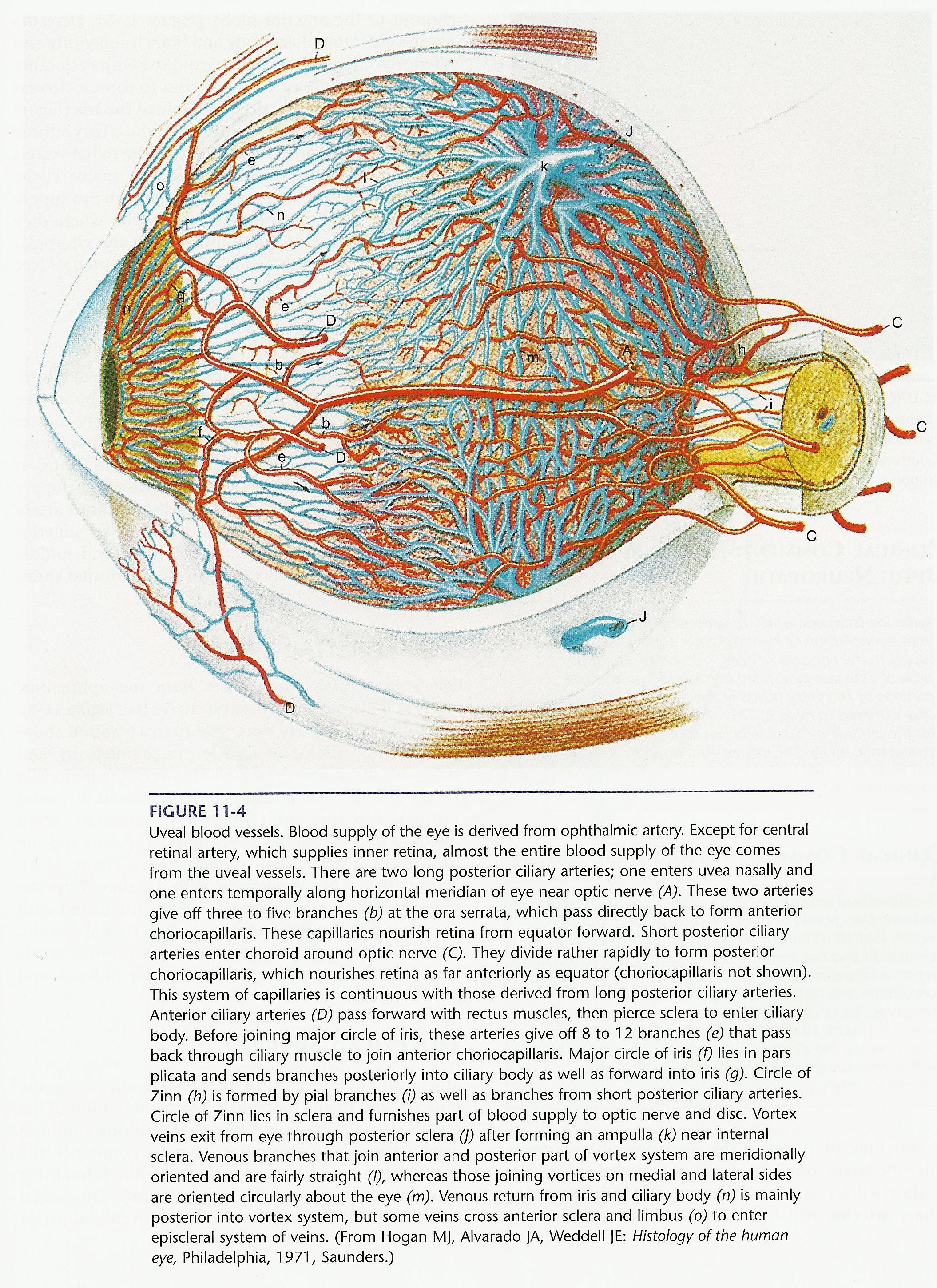
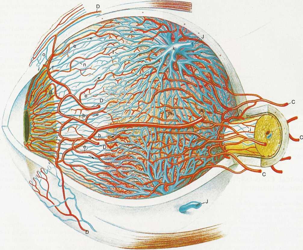
Uveal blood vessels. Blood supply of the eye is derived from ophthalmic artery. Except for central retinal artery, which supplies inner retina, almost the entire blood supply of the eye comes from the uveal vessels. There are two long posterior ciliary arteries; one enters uvea nasally and one enters temporally along horizontal meridian of eye near optic nerve (A). These two arteries give off three to five branches (b) at the ora serrata, which pass directly back to form anterior choriocapillaris. These capillaries nourish retina from equator forward. Short posterior ciliary arteries enter choroid around optic nerve (C). They divide rather rapidly to form posterior choriocapillaris, which nourishes retina as far anteriorly as equator (choriocapillaris not shown). This system of capillaries is continuous with those derived from long posterior ciliary arteries. Anterior ciliary arteries (D) pass forward with rectus muscles, then pierce selera to enter ciliary body. Before joining major circle of iris, these arteries give off 8 to 12 branches (e) that pass back through ciliary muscle to join anterior choriocapillaris. Major circle of iris (f) lies in pars plicata and sends branches posteriorly into ciliary body as well as forward into iris (g). Circle of Zinn (h) is formed by piał branches (i) as well as branches from short posterior ciliary arteries. Circle of Zinn lies in selera and furnishes part of blood supply to optic nerve and disc. Vortex veins exit from eye through posterior selera (]) after forming an ampulla (k) near internal selera. Venous branches that join anterior and posterior part of vortex system are meridionally oriented and are fairly straight (I), whereas those joining vortices on medial and lateral sides are oriented circularly about the eye (m). Venous return from iris and ciliary body (n) is mainly posterior into vortex system, but some veins cross anterior selera and limbus (o) to enter episcleral system of veins. (From Hogan MJ, Alvarado JA, Weddell JE: Histology of the human eye, Philadelphia, 1971, Saunders.)
Wyszukiwarka
Podobne podstrony:
SCAN0150 crop FIGURĘ 11-9 Branches of external carotid artery that supply ocular adnexa. (Redrawn fr
SCAN0152 crop FIGURĘ 12-1 Orbit viewed from above showing branches of ophthalmic nerve.
8 11 Figurę 8-11 Stripping and cross-fiber stroking of iliacus with fingertips (A), supported thumb
SCAN0042 crop FIGURĘ 2-1 Corneal dimensions. A, Radius of curvature of cornea and selera. B, View fr
Figurę 12: The components of the KASUMI błock cipher (from [6]). • Encryption key
SCAN0033 crop Eye Axes Since the eye is not rotationally symmetric (i.e. the centers of curvature of
11 11 2 T topie explain that the topie of the speech is is important to the audience.
SCAN0033 crop Eye Axes Since the eye is not rotationally symmetric (i.e. the centers of curvature of
140 Kostas P. Kyrris 8 11. Though the end of the story is the liberation of t
49 (253) 92 The Viking Age in Denmark Figurę 25 Reconstructed tcnth-century pit-houses of the town o
więcej podobnych podstron