skanuj0016 (266)
Neuroanatomy 10. Sectional Anatomy of the Brain
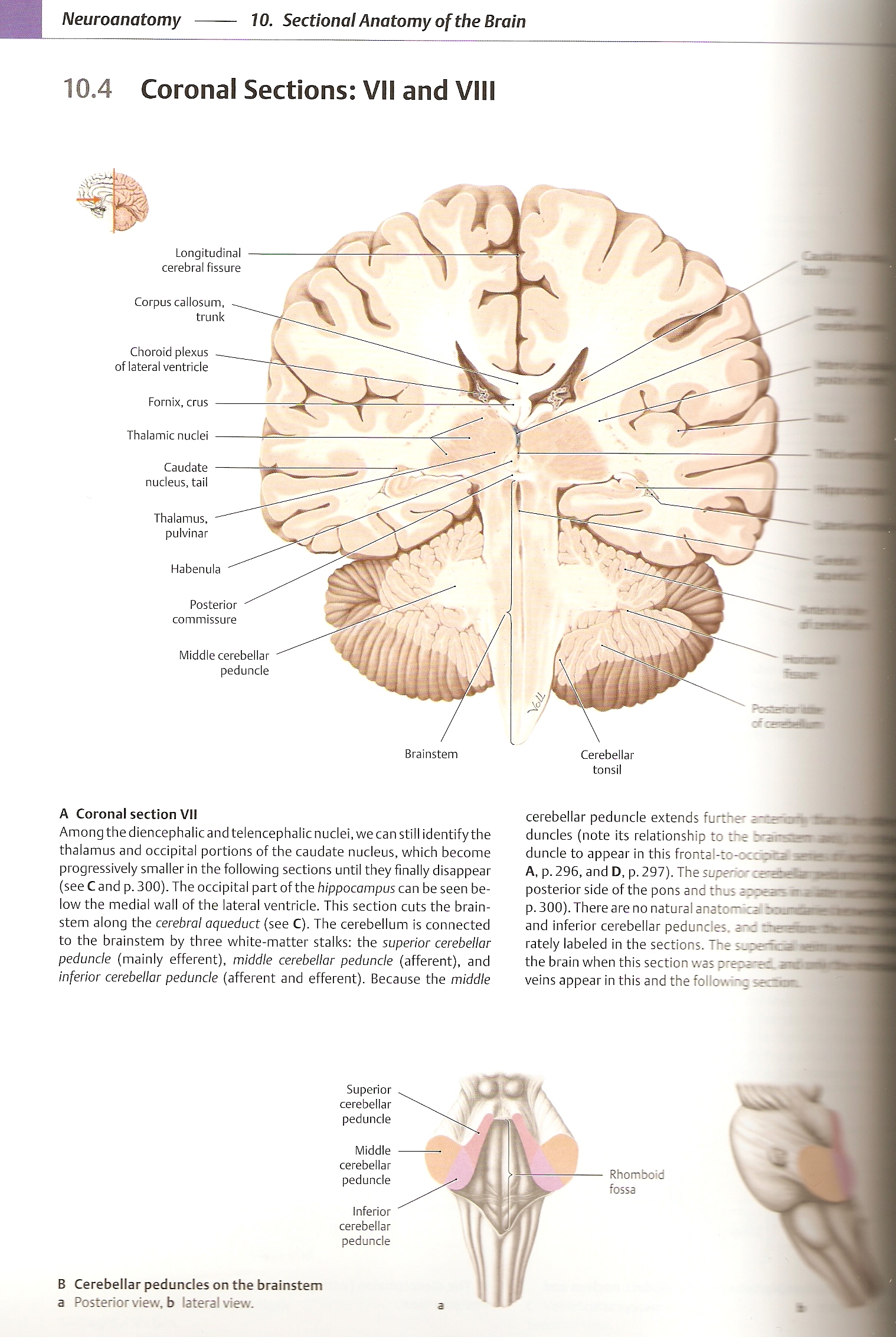
10.4 Coronal Sections: VII and VIII
Neuroanatomy 10. Sectional Anatomy of the Brain
Longitudinal cerebral fissure
Corpus callosum, trunk
Choroid plexus of lateral ventricle
Fornix, crus Thalamic nudei
Caudate nudeus, taił
Thalamus,
pulvinar
Habenula
Posterior
commissure
Brainstem
Cerebellar
tonsil
Middle cerebellar peduncle
A Coronal section VII
Among the diencephalic and telencephalic nudei, we can still identify the thalamus and ocdpital portions of the caudate nudeus, which become progressively smaller in the following sections until they finally disappear (see C and p. 300). The occipital part of the hippocampus can be seen be-low the medial wali of the lateral ventricle. This section cuts the brainstem along the cerebral aqueduct (see C). The cerebellum is connected to the brainstem by three white-matter stalks: the superior cerebellar peduncle (mainly efferent), middle cerebellar peduncle (afferent), and inferior cerebellar peduncle (afferent and efferent). Because the middle
Superior
cerebellar
peduncle
Middle
cerebellar
peduncle
Inferior
cerebellar
peduncle
B Cerebellar pedundes on the brainstem a Posterior view. b lateral view.
cerebellar peduncle extends further i ■ r o~, duncles (notę its relationship to the braiisa duncle to appear in this frontal-to-occor= a A, p. 296, and D, p. 297). The superior csM posterior side of the pons and thus aopeasia p. 300). There are no natural anatom ca roana and inferior cerebellar pedundes. and rual rately labeled in the sections. The the brain when this section was prera-ec. m veins appear in this and the followirę ^“or.
Rhomboid
fossa
Wyszukiwarka
Podobne podstrony:
skanuj0016 (266) Neuroanatomy 10. Sectional Anatomy of the Brain10.4 Coronal Sections: VII and VIII
skanuj0015 (279) Neuroanatomy 10. Sectional Anatomy of the Brain -landtudinal Mrafcsure ■PHlfillilll
88514 skanuj0015 (279) Neuroanatomy 10. Sectional Anatomy of the Brain -landtudinal Mrafcsure ■PHlfi
SCAN0131 96 Clinical Anatomy of the Visual System 96 Clinical Anatomy of the Visual System FIGURĘ 5-
SCAN0145 178 Clinical Anatomy of the Visual System Epimysium Perimysium EndomysiumFIGURĘ 10-1 Connec
SCAN0131 96 Clinical Anatomy of the Visual System 96 Clinical Anatomy of the Visual System FIGURĘ 5-
10 1 Fascia lata Medial collateral ligament Patellar ligament Extensor retinacula Figurę 10-1 Anatom
75024 SCAN0131 96 Clinical Anatomy of the Visual System 96 Clinical Anatomy of the Visual System FIG
10 7 Figurę 10-7 Anatomy of the plantar fascia
SCAN0042 10 Clinical Anatomy of the Visual System 10 Clinical Anatomy of the Visual System FIGURĘ 2-
SCAN0145 178 Clinical Anatomy of the Visual System Epimysium Perimysium EndomysiumFIGURĘ 10-1 Connec
skanuj0059 (23) 180 MARTA DEREK objectives of the economic development of the commune. For example,
więcej podobnych podstron